A DICOM File Is Not a Patient Experience
Giving patients access to medical images is important, but a folder of DICOM files does not by itself provide understanding, context, or a clear next step.
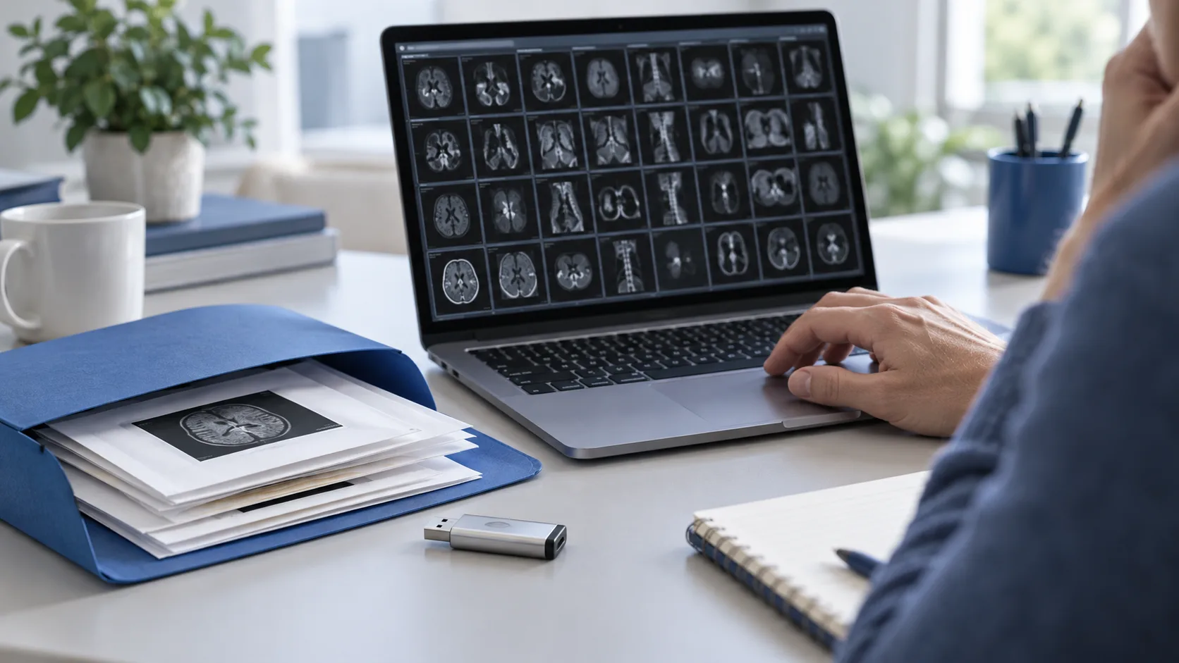
Patients are increasingly able to obtain copies of their medical images. Sometimes the files arrive on a disc, USB drive, download link, or patient portal. The format is often DICOM, the standard used to store medical images together with technical and identifying metadata.
That access matters. But access to a file is not the same as access to understanding.
What a patient receives
A CT or MRI study may contain hundreds or thousands of individual images. They may be grouped into multiple series, orientations, reconstructions, and contrast phases. The folder can also contain metadata such as the study date, body region, modality, institution, patient identifier, and acquisition settings.
For a radiologist, this structure is part of a professional workflow. For a patient, it can look like an unintelligible collection of files with unfamiliar names.
Opening a single image may not show the relevant slice. Looking at a different series may create a misleading impression. A screenshot without its context can be difficult to interpret and easy to mislabel.
Viewing is not interpreting
A DICOM viewer can help someone inspect images, scroll through slices, adjust window and level, and understand how a study is organized. Those functions are useful for transparency and for preparing questions.
They do not turn a patient into a radiologist.
Image interpretation depends on the complete study, image quality, clinical history, comparison examinations, and professional training. A viewer should therefore make its purpose clear: it helps people access and navigate their files; it is not a regulated diagnostic workstation and it does not replace formal review.
The privacy problem inside the file
DICOM files can contain more than pixels. Depending on the source, metadata may include a patient name, patient ID, date of birth, institution, study date, and other identifiers. A person may share an image online without realizing that identifying information travels with it.
Patient-friendly tools should explain this before a user exports, uploads, or shares a study. Privacy guidance should be practical:
- Check the destination before uploading files.
- Avoid sharing a full study publicly.
- Remove or anonymize identifiers when appropriate.
- Remember that screenshots may still reveal clinical information.
- Ask the imaging provider how to request a de-identified copy.
The missing layer is context
Patients do not just need a way to open an image. They need help connecting the image to the report, the examination date, the body region, and the questions they want to ask their doctor.
Useful context might include:
- Which series is being viewed;
- Whether the study is complete or only a selected export;
- Which findings are described in the accompanying report;
- What the report recommends discussing;
- Which questions cannot be answered from the files alone.
This is where education can sit alongside access. A tool can explain the difference between a series and a slice, define common terms, and help a patient prepare for a clinical conversation without pretending to provide a diagnosis.
Design for the moment after download
The real patient experience often begins after the files leave the hospital. The person may be anxious, alone, and looking for an explanation before the follow-up appointment. A responsible product should meet that moment with clear boundaries rather than a confident interpretation.
The goal is not to make every patient an imaging expert. It is to prevent a folder of opaque files from becoming a dead end.
Medical transparency is strongest when people can access their data, understand what it contains, protect their privacy, and know which questions belong with a qualified clinician.